


Microcystin Plate Kit
I. Principle
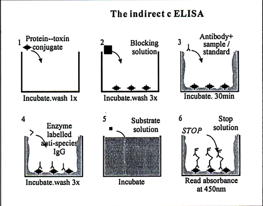
ELISA is a form of immunoenzymatic technology that combines the specificity of antigen–antibody reactions with the sensitivity of enzyme reactions. The principle is that an enzyme molecule is bound to an antibody (or antigen) to form a stable enzyme-labelled conjugate. When this conjugate binds to the corresponding antigen (or antibody) on a solid support, a visible colour reaction is produced in the presence of substrate; the colour intensity is proportional to the amount of antigen or antibody, and the absorbance can be measured with an ELISA reader for quantitative analysis. The method is specific, sensitive, objective, simple and safe, and is now widely used not only in microbiology but also in many other fields. The working principle of this kit is shown in the figure below.
II. Reagents supplied
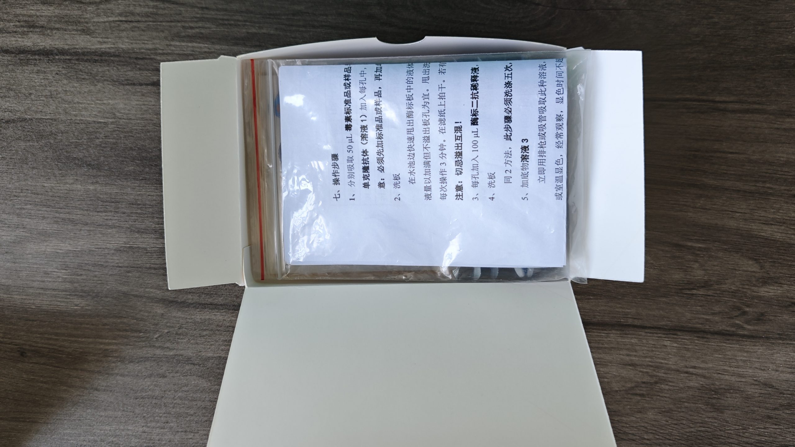
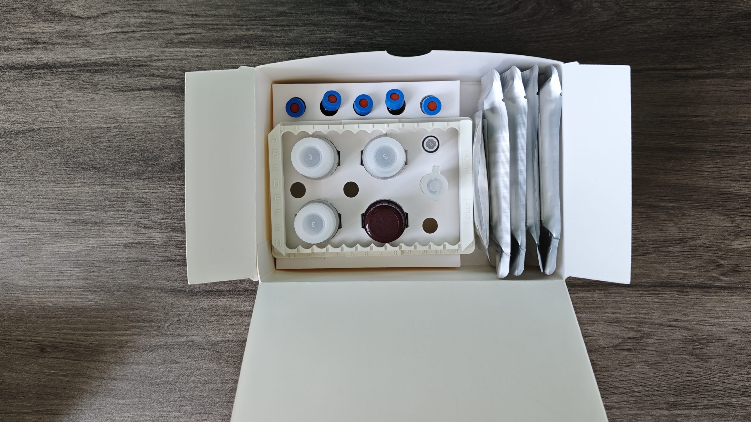
The ELISA kit contains the following items:
1. A pre-coated 96-well microplate
2. Standard 1: 0.1 ng/mL MC-LR standard solution
3. Standard 2: 0.2 ng/mL MC-LR standard solution
4. Standard 3: 0.5 ng/mL MC-LR standard solution
5. Standard 4: 1 ng/mL MC-LR standard solution
6. Standard 5: 2 ng/mL MC-LR standard solution
7. 1 bottle of Solution 1 (monoclonal antibody)
8. 1 vial of Solution 2 (this solution is only for diluting the secondary antibody, not the secondary-antibody solution itself; measure according to the dilution ratio!)
9. Secondary antibody (the small tube contains 2.4 μL of enzyme-labelled secondary antibody; store at 4 °C).
10. 1 bottle of Solution 3 (substrate)
11. 1 bottle of Solution 4 (stop solution)
12. 1 bag of solid PBS (for preparing wash buffer)
13. 1 vial of Tween 20 (for preparing wash buffer)
III. Equipment and reagents required
In addition to the contents of the kit, the following equipment and reagents are required:
1. Microplate reader (and a plate washer if available)
2. Micropipette (100 μL) (if possible, an 8- or 12-channel micropipette)
3. Pipettes and rubber bulbs (for plate washing; not needed if a plate washer is available)
4. Purified water (for preparing wash buffer)
5. Glass bottle (to hold wash buffer)
6. Timer (to control the time of each step accurately)
7. Sealing film (to prevent evaporation from the wells)
IV. Preparation of the enzyme-labelled secondary antibody
Dilute the secondary antibody in the small tube 1:5,000 with Solution 2, dissolve and mix thoroughly, and prepare immediately before use.
V. Preparation of wash buffer
Make up the solid PBS to 1 L with purified water (pH 7.4–7.6). Remove 0.5 ml from the small plastic tube
Tween 20 into the PBS solution prepared above. Store at room temperature.
VI. Sample pretreatment
1. Treatment of algal samples: collected samples may be processed in three ways: a. freeze–thaw (2–3 cycles); b. ultrasonication
c. Disruption with a cell press.
2. Treatment of water samples: centrifuge the collected water and take the supernatant, or filter.
3. Treatment of aquatic products: the method is relatively complex; see the literature method: Valéria Freitas de Magalhães,
Raquel Moraes Soares, Sandra M.F.O.Azevedo. Toxicon, 2001,39:1077
VII. Procedure
1. Pipette 50 μL of toxin standard or sample into the corresponding wells (see Table 1), then pipette 50 μL
of monoclonal antibody (Solution 1) to each well and mix gently. Incubate at 37 °C or room temperature for 90 minutes.
Note: standards or samples must be added first, then the monoclonal antibody solution.
2. Wash the plate
Quickly shake the liquid out of the microplate over a sink. Fill each well with wash buffer without overflowing, then empty and repeat this wash three times, 3 minutes each time. Blot dry on filter paper. If a plate washer is available, it may be used instead.
Note: do not overflow or mix wells!
3. Add 100 μL of diluted enzyme-labelled secondary antibody to each well and incubate at 37 °C or room temperature for 30 minutes.
4. Wash the plate
As in step 2, this step must be washed five times, 3 minutes each.
5. Add substrate solution 3
Immediately add 100 µl of this solution to each well with a multichannel or single pipette. Develop at 37 °C or room temperature, watching frequently; do not exceed 30 minutes.
6. Stop the reaction
When a clear colour has developed, immediately add 50 μL of Solution 4 (1 mol/L H2SO4) to stop the reaction.
7. Interpret the results
By visual inspection (white background), the negative-control wells should be clearly yellow.
Measure optical density with the microplate reader at λ = 450 nm. Note: measure within 30 minutes after adding stop solution.
Note: pipette volumes accurately, and do not allow reagent to flow from one well to another. Use a separate pipette (or tip) for each reagent.
VIII. Data processing
1. Layout of additions to the corresponding wells of the plate:
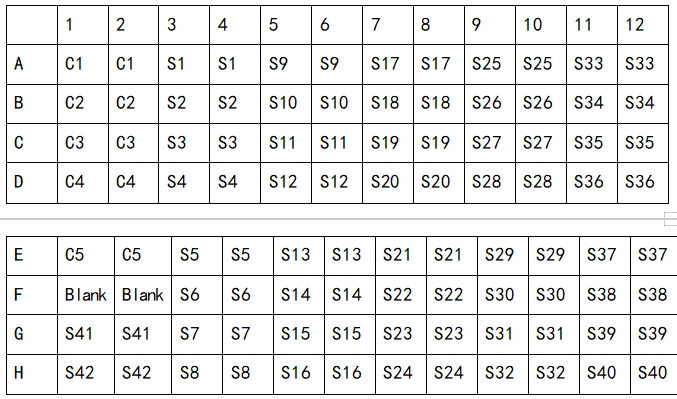
C1, C2, C3, C4, C5: toxin standards; S1–S42: samples to be tested
2. Calculate the mean OD450 of each standard or sample and compute B0 by the following formula.
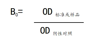
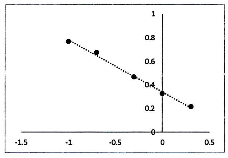
3. Construct a standard curve from the relationship between the B0 values of the standards and the logarithms of the corresponding concentrations. The standard curve in the figure below
The curve may be used as a reference.
4. Calculate the concentration of each sample from the standard curve.
Development of this kit was supported by the National High-tech R&D Program (863 Program) projects “Technical system for cyanotoxin analysis and rapid on-site testing” (2001AA641030) and “Rapid on-site cyanotoxin testing technology and industrialization” (2003AA641020).



Reviews
There are no reviews yet.